What Structure Allows a Bone to Grow in Length?
Os Tissue and the Skeletal System
Os Structure
Learning Objectives
By the end of this section, y'all volition exist able to:
- Identify the anatomical features of a bone
- Define and list examples of bone markings
- Depict the histology of os tissue
- Compare and contrast meaty and spongy bone
- Identify the structures that etch compact and spongy bone
- Describe how bones are nourished and innervated
Os tissue (osseous tissue) differs greatly from other tissues in the body. Bone is hard and many of its functions depend on that feature hardness. Afterward discussions in this chapter will show that os is likewise dynamic in that its shape adjusts to arrange stresses. This section will examine the gross anatomy of bone kickoff and and then move on to its histology.
Gross Anatomy of Bone
The construction of a long bone allows for the best visualization of all of the parts of a bone ((Figure)). A long bone has two parts: the diaphysis and the epiphysis. The diaphysis is the tubular shaft that runs between the proximal and distal ends of the bone. The hollow region in the diaphysis is chosen the medullary cavity, which is filled with yellowish marrow. The walls of the diaphysis are composed of dumbo and hard compact bone.
Beefcake of a Long Bone
A typical long bone shows the gross anatomical characteristics of bone.
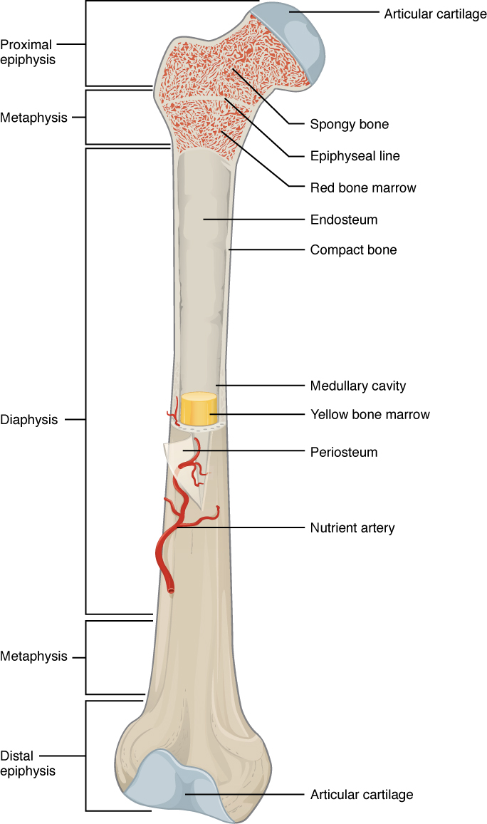
The wider section at each terminate of the os is called the epiphysis (plural = epiphyses), which is filled with spongy bone. Ruddy marrow fills the spaces in the spongy bone. Each epiphysis meets the diaphysis at the metaphysis, the narrow area that contains the epiphyseal plate (growth plate), a layer of hyaline (transparent) cartilage in a growing bone. When the bone stops growing in early adulthood (approximately xviii–21 years), the cartilage is replaced past osseous tissue and the epiphyseal plate becomes an epiphyseal line.
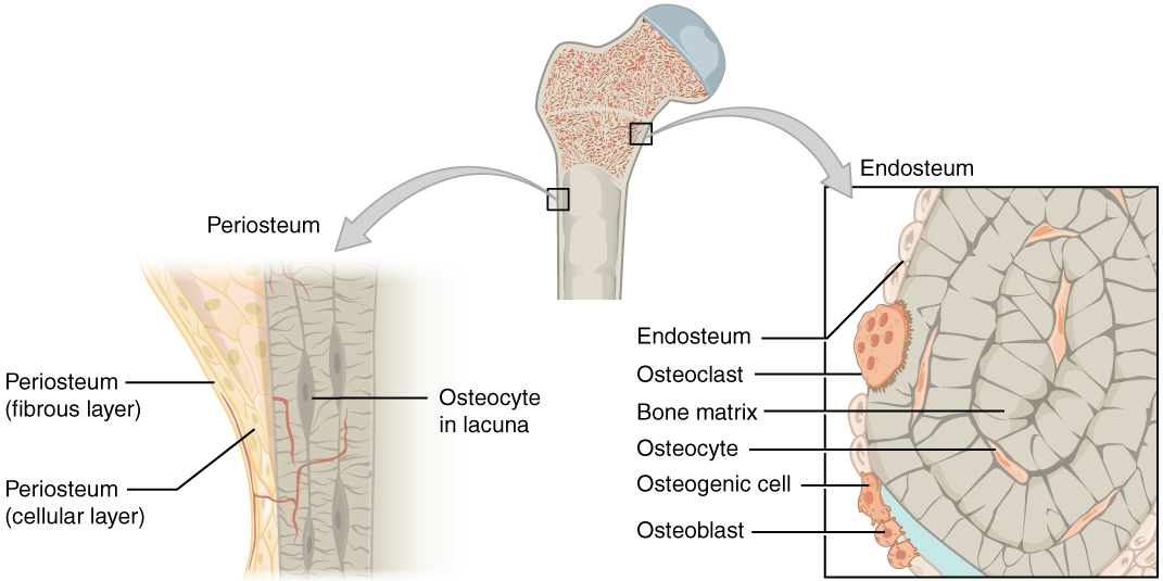
The medullary cavity has a delicate membranous lining chosen the endosteum (finish- = "inside"; oste- = "os"), where bone growth, repair, and remodeling occur. The outer surface of the os is covered with a fibrous membrane chosen the periosteum (peri– = "around" or "surrounding"). The periosteum contains blood vessels, nerves, and lymphatic vessels that nourish compact bone. Tendons and ligaments too adhere to bones at the periosteum. The periosteum covers the entire outer surface except where the epiphyses meet other basic to form joints ((Figure)). In this region, the epiphyses are covered with articular cartilage, a thin layer of cartilage that reduces friction and acts as a shock absorber.
Periosteum and Endosteum
The periosteum forms the outer surface of bone, and the endosteum lines the medullary cavity.
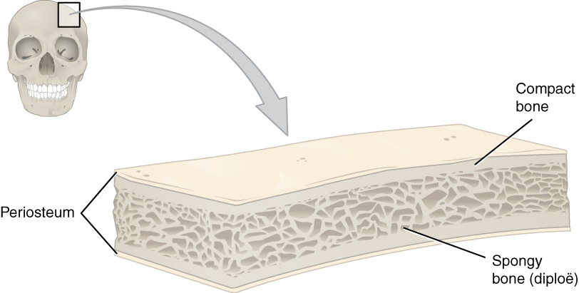
Flat basic, like those of the cranium, consist of a layer of diploë (spongy bone), lined on either side by a layer of compact os ((Effigy)). The two layers of compact bone and the interior spongy bone work together to protect the internal organs. If the outer layer of a cranial bone fractures, the encephalon is even so protected by the intact inner layer.
Beefcake of a Apartment Bone
This cross-section of a apartment os shows the spongy bone (diploë) lined on either side past a layer of compact bone.
Bone Markings
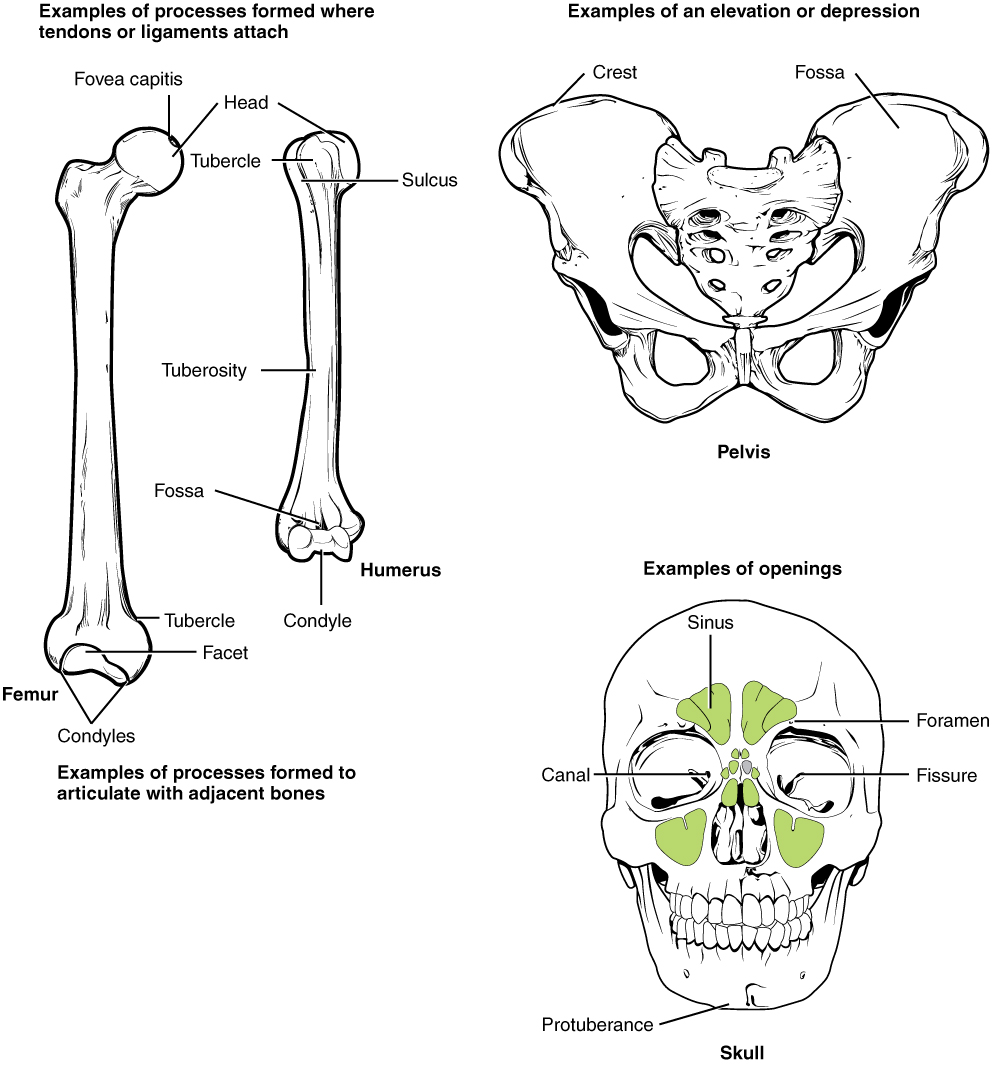
The surface features of bones vary considerably, depending on the part and location in the body. (Effigy) describes the bone markings, which are illustrated in ((Figure)). There are three general classes of bone markings: (ane) articulations, (2) projections, and (3) holes. As the name implies, an articulation is where two bone surfaces come together (articulus = "joint"). These surfaces tend to conform to i another, such as one existence rounded and the other cupped, to facilitate the part of the articulation. A projection is an area of a bone that projects above the surface of the bone. These are the attachment points for tendons and ligaments. In full general, their size and shape is an indication of the forces exerted through the zipper to the bone. A hole is an opening or groove in the os that allows blood vessels and nerves to enter the bone. As with the other markings, their size and shape reflect the size of the vessels and nerves that penetrate the os at these points.
| Os Markings | ||
|---|---|---|
| Marking | Description | Instance |
| Articulations | Where two bones meet | Knee joint |
| Head | Prominent rounded surface | Head of femur |
| Facet | Apartment surface | Vertebrae |
| Condyle | Rounded surface | Occipital condyles |
| Projections | Raised markings | Spinous process of the vertebrae |
| Protuberance | Protruding | Chin |
| Procedure | Prominence feature | Transverse process of vertebra |
| Spine | Sharp process | Ischial spine |
| Tubercle | Small, rounded procedure | Tubercle of humerus |
| Tuberosity | Crude surface | Deltoid tuberosity |
| Line | Slight, elongated ridge | Temporal lines of the parietal basic |
| Crest | Ridge | Iliac crest |
| Holes | Holes and depressions | Foramen (holes through which blood vessels can pass through) |
| Fossa | Elongated basin | Mandibular fossa |
| Fovea | Small pit | Fovea capitis on the head of the femur |
| Sulcus | Groove | Sigmoid sulcus of the temporal bones |
| Culvert | Passage in bone | Auditory canal |
| Fissure | Slit through bone | Auricular fissure |
| Foramen | Hole through os | Foramen magnum in the occipital os |
| Meatus | Opening into culvert | External auditory meatus |
| Sinus | Air-filled space in bone | Nasal sinus |
Bone Features
The surface features of bones depend on their part, location, attachment of ligaments and tendons, or the penetration of blood vessels and nerves.
Bone Cells and Tissue
Bone contains a relatively modest number of cells entrenched in a matrix of collagen fibers that provide a surface for inorganic salt crystals to adhere. These table salt crystals form when calcium phosphate and calcium carbonate combine to create hydroxyapatite, which incorporates other inorganic salts like magnesium hydroxide, fluoride, and sulfate as it crystallizes, or calcifies, on the collagen fibers. The hydroxyapatite crystals requite bones their hardness and force, while the collagen fibers give them flexibility so that they are not brittle.
Although os cells etch a small corporeality of the os volume, they are crucial to the part of basic. 4 types of cells are found within bone tissue: osteoblasts, osteocytes, osteogenic cells, and osteoclasts ((Figure)).
Bone Cells
Iv types of cells are found within bone tissue. Osteogenic cells are undifferentiated and develop into osteoblasts. When osteoblasts get trapped within the calcified matrix, their structure and office changes, and they go osteocytes. Osteoclasts develop from monocytes and macrophages and differ in appearance from other bone cells.

The osteoblast is the os cell responsible for forming new bone and is found in the growing portions of os, including the periosteum and endosteum. Osteoblasts, which exercise not divide, synthesize and secrete the collagen matrix and calcium salts. As the secreted matrix surrounding the osteoblast calcifies, the osteoblast become trapped within it; as a outcome, information technology changes in structure and becomes an osteocyte, the primary prison cell of mature os and the most common blazon of bone cell. Each osteocyte is located in a space called a lacuna and is surrounded by bone tissue. Osteocytes maintain the mineral concentration of the matrix via the secretion of enzymes. Like osteoblasts, osteocytes lack mitotic activity. They can communicate with each other and receive nutrients via long cytoplasmic processes that extend through canaliculi (atypical = canaliculus), channels within the bone matrix.
If osteoblasts and osteocytes are incapable of mitosis, then how are they replenished when onetime ones die? The respond lies in the properties of a third category of os cells—the osteogenic cell. These osteogenic cells are undifferentiated with loftier mitotic action and they are the simply bone cells that divide. Immature osteogenic cells are plant in the deep layers of the periosteum and the marrow. They differentiate and develop into osteoblasts.
The dynamic nature of bone means that new tissue is constantly formed, and old, injured, or unnecessary bone is dissolved for repair or for calcium release. The prison cell responsible for bone resorption, or breakup, is the osteoclast. They are found on bone surfaces, are multinucleated, and originate from monocytes and macrophages, ii types of white blood cells, not from osteogenic cells. Osteoclasts are continually breaking down old bone while osteoblasts are continually forming new bone. The ongoing balance between osteoblasts and osteoclasts is responsible for the constant but subtle reshaping of bone. (Figure) reviews the os cells, their functions, and locations.
| Bone Cells | ||
|---|---|---|
| Prison cell type | Function | Location |
| Osteogenic cells | Develop into osteoblasts | Deep layers of the periosteum and the marrow |
| Osteoblasts | Os formation | Growing portions of os, including periosteum and endosteum |
| Osteocytes | Maintain mineral concentration of matrix | Entrapped in matrix |
| Osteoclasts | Os resorption | Bone surfaces and at sites of sometime, injured, or unneeded bone |
Meaty and Spongy Os
The differences between compact and spongy bone are all-time explored via their histology. Most bones incorporate meaty and spongy osseous tissue, but their distribution and concentration vary based on the bone's overall part. Compact bone is dumbo so that it tin withstand compressive forces, while spongy (cancellous) bone has open spaces and supports shifts in weight distribution.
Compact Bone
Meaty bone is the denser, stronger of the two types of bone tissue ((Figure)). It can be establish under the periosteum and in the diaphyses of long bones, where information technology provides support and protection.
Diagram of Compact Bone
(a) This cross-sectional view of compact bone shows the basic structural unit, the osteon. (b) In this micrograph of the osteon, you lot can clearly come across the concentric lamellae and key canals. LM × 40. (Micrograph provided by the Regents of University of Michigan Medical School © 2012)
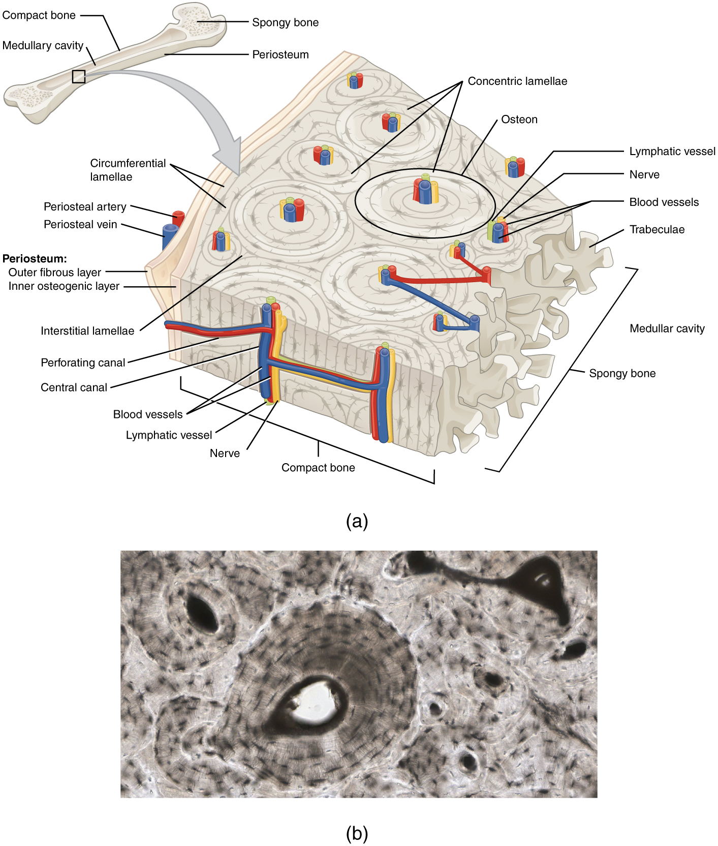
The microscopic structural unit of compact bone is called an osteon, or Haversian arrangement. Each osteon is composed of concentric rings of calcified matrix called lamellae (atypical = lamella). Running down the center of each osteon is the key canal, or Haversian canal, which contains claret vessels, nerves, and lymphatic vessels. These vessels and nerves branch off at right angles through a perforating canal, also known as Volkmann'south canals, to extend to the periosteum and endosteum.
The osteocytes are located within spaces called lacunae (singular = lacuna), found at the borders of adjacent lamellae. As described earlier, canaliculi connect with the canaliculi of other lacunae and somewhen with the cardinal canal. This system allows nutrients to be transported to the osteocytes and wastes to exist removed from them.
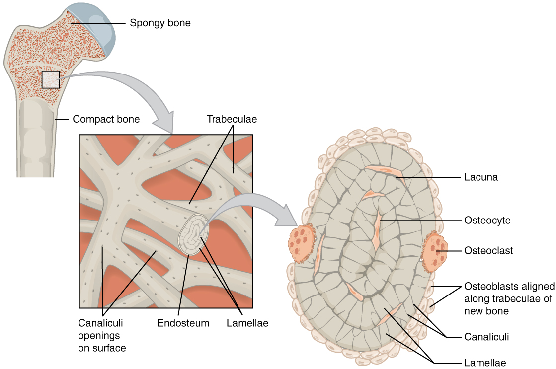
Spongy (Cancellous) Bone
Like compact bone, spongy os, also known as cancellous bone, contains osteocytes housed in lacunae, but they are not bundled in concentric circles. Instead, the lacunae and osteocytes are found in a lattice-like network of matrix spikes called trabeculae (singular = trabecula) ((Figure)). The trabeculae may appear to be a random network, but each trabecula forms along lines of stress to provide strength to the os. The spaces of the trabeculated network provide balance to the dense and heavy compact os by making bones lighter so that muscles can movement them more easily. In addition, the spaces in some spongy bones contain red marrow, protected by the trabeculae, where hematopoiesis occurs.
Diagram of Spongy Bone
Spongy bone is equanimous of trabeculae that contain the osteocytes. Red marrow fills the spaces in some bones.
Crumbling and the…
Skeletal System: Paget's Disease Paget's disease commonly occurs in adults over age 40. It is a disorder of the bone remodeling process that begins with overactive osteoclasts. This means more than bone is resorbed than is laid down. The osteoblasts attempt to compensate but the new bone they lay down is weak and brittle and therefore prone to fracture.
While some people with Paget'due south illness accept no symptoms, others experience pain, bone fractures, and bone deformities ((Figure)). Basic of the pelvis, skull, spine, and legs are the most commonly affected. When occurring in the skull, Paget's disease can cause headaches and hearing loss.
Paget's Disease
Normal leg bones are relatively straight, but those afflicted past Paget's disease are porous and curved.
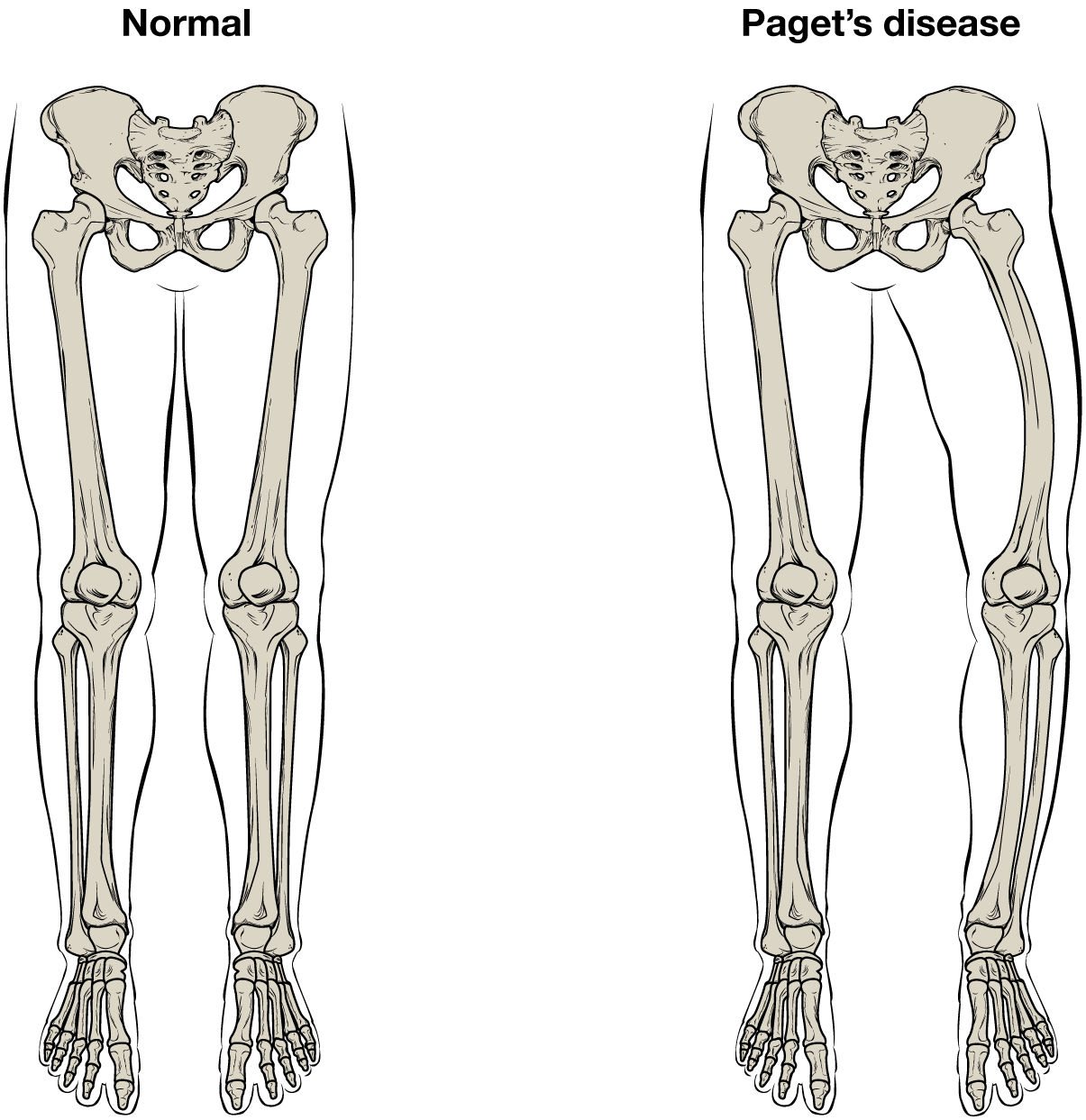
What causes the osteoclasts to become overactive? The answer is still unknown, merely hereditary factors seem to play a role. Some scientists believe Paget's disease is due to an as-yet-unidentified virus.
Paget's disease is diagnosed via imaging studies and lab tests. Ten-rays may show os deformities or areas of bone resorption. Bone scans are too useful. In these studies, a dye containing a radioactive ion is injected into the body. Areas of os resorption have an affinity for the ion, and then they volition low-cal upward on the scan if the ions are absorbed. In addition, blood levels of an enzyme chosen alkaline phosphatase are typically elevated in people with Paget's disease.
Bisphosphonates, drugs that decrease the activity of osteoclasts, are oftentimes used in the treatment of Paget's affliction. Nevertheless, in a small percentage of cases, bisphosphonates themselves accept been linked to an increased run a risk of fractures because the old bone that is left later bisphosphonates are administered becomes worn out and breakable. Yet, virtually doctors feel that the benefits of bisphosphonates more than than outweigh the gamble; the medical professional has to weigh the benefits and risks on a case-past-instance basis. Bisphosphonate treatment can reduce the overall run a risk of deformities or fractures, which in turn reduces the chance of surgical repair and its associated risks and complications.
Blood and Nerve Supply
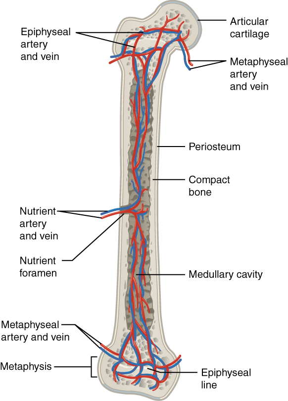
The spongy bone and medullary cavity receive nourishment from arteries that pass through the compact os. The arteries enter through the nutrient foramen (plural = foramina), pocket-sized openings in the diaphysis ((Figure)). The osteocytes in spongy bone are nourished past blood vessels of the periosteum that penetrate spongy bone and blood that circulates in the marrow cavities. As the blood passes through the marrow cavities, it is nerveless by veins, which so pass out of the bone through the foramina.
In addition to the claret vessels, nerves follow the same paths into the bone where they tend to concentrate in the more metabolically active regions of the bone. The nerves sense hurting, and it appears the fretfulness also play roles in regulating blood supplies and in bone growth, hence their concentrations in metabolically agile sites of the os.
Diagram of Blood and Nerve Supply to Bone
Blood vessels and nerves enter the os through the food foramen.
Watch this video to see the microscopic features of a bone.
Chapter Review
A hollow medullary cavity filled with yellow marrow runs the length of the diaphysis of a long bone. The walls of the diaphysis are meaty bone. The epiphyses, which are wider sections at each cease of a long bone, are filled with spongy os and red marrow. The epiphyseal plate, a layer of hyaline cartilage, is replaced by osseous tissue equally the organ grows in length. The medullary cavity has a delicate membranous lining called the endosteum. The outer surface of os, except in regions covered with articular cartilage, is covered with a fibrous membrane called the periosteum. Flat bones consist of 2 layers of compact bone surrounding a layer of spongy bone. Bone markings depend on the function and location of bones. Articulations are places where ii bones see. Projections stick out from the surface of the os and provide zipper points for tendons and ligaments. Holes are openings or depressions in the bones.
Bone matrix consists of collagen fibers and organic footing substance, primarily hydroxyapatite formed from calcium salts. Osteogenic cells develop into osteoblasts. Osteoblasts are cells that make new bone. They become osteocytes, the cells of mature os, when they get trapped in the matrix. Osteoclasts engage in bone resorption. Compact os is dense and composed of osteons, while spongy bone is less dense and made up of trabeculae. Blood vessels and nerves enter the bone through the nutrient foramina to nourish and innervate bones.
Review Questions
Which of the following occurs in the spongy os of the epiphysis?
- bone growth
- os remodeling
- hematopoiesis
- shock absorption
The diaphysis contains ________.
- the metaphysis
- fatty stores
- spongy os
- compact bone
The fibrous membrane covering the outer surface of the bone is the ________.
- periosteum
- epiphysis
- endosteum
- diaphysis
Which of the following are incapable of undergoing mitosis?
- osteoblasts and osteoclasts
- osteocytes and osteoclasts
- osteoblasts and osteocytes
- osteogenic cells and osteoclasts
Which cells do not originate from osteogenic cells?
- osteoblasts
- osteoclasts
- osteocytes
- osteoprogenitor cells
Which of the following are plant in compact bone and cancellous os?
- Haversian systems
- Haversian canals
- lamellae
- lacunae
Which of the following are merely institute in cancellous os?
- canaliculi
- Volkmann's canals
- trabeculae
- calcium salts
The area of a bone where the nutrient foramen passes forms what kind of os marker?
- a hole
- a facet
- a culvert
- a fissure
Disquisitional Thinking Questions
If the articular cartilage at the end of one of your long basic were to degenerate, what symptoms practice you think you would experience? Why?
If the articular cartilage at the end of one of your long bones were to deteriorate, which is really what happens in osteoarthritis, you would experience joint pain at the end of that bone and limitation of motion at that joint because there would be no cartilage to reduce friction between adjacent basic and in that location would be no cartilage to act as a shock cushion.
In what ways is the structural makeup of meaty and spongy bone well suited to their respective functions?
The densely packed concentric rings of matrix in meaty bone are ideal for resisting compressive forces, which is the part of compact bone. The open up spaces of the trabeculated network of spongy bone allow spongy bone to support shifts in weight distribution, which is the function of spongy bone.
Glossary
- articular cartilage
- thin layer of cartilage covering an epiphysis; reduces friction and acts equally a stupor absorber
- articulation
- where two bone surfaces meet
- canaliculi
- (singular = canaliculus) channels within the os matrix that firm one of an osteocyte'southward many cytoplasmic extensions that information technology uses to communicate and receive nutrients
- cardinal canal
- longitudinal channel in the centre of each osteon; contains blood vessels, fretfulness, and lymphatic vessels; likewise known as the Haversian canal
- compact os
- dense osseous tissue that tin withstand compressive forces
- diaphysis
- tubular shaft that runs between the proximal and distal ends of a long bone
- diploë
- layer of spongy os, that is sandwiched between two the layers of compact bone found in flat bones
- endosteum
- delicate bleary lining of a bone's medullary cavity
- epiphyseal plate
- (likewise, growth plate) sheet of hyaline cartilage in the metaphysis of an young os; replaced past bone tissue as the organ grows in length
- epiphysis
- broad section at each end of a long bone; filled with spongy os and red marrow
- hole
- opening or depression in a bone
- lacunae
- (singular = lacuna) spaces in a bone that firm an osteocyte
- medullary cavity
- hollow region of the diaphysis; filled with xanthous marrow
- nutrient foramen
- pocket-sized opening in the middle of the external surface of the diaphysis, through which an artery enters the bone to provide nourishment
- osteoblast
- cell responsible for forming new bone
- osteoclast
- cell responsible for resorbing bone
- osteocyte
- primary prison cell in mature os; responsible for maintaining the matrix
- osteogenic prison cell
- undifferentiated jail cell with high mitotic activity; the just os cells that separate; they differentiate and develop into osteoblasts
- osteon
- (also, Haversian system) bones structural unit of meaty bone; made of concentric layers of calcified matrix
- perforating canal
- (also, Volkmann's culvert) channel that branches off from the key culvert and houses vessels and nerves that extend to the periosteum and endosteum
- periosteum
- fibrous membrane covering the outer surface of bone and continuous with ligaments
- projection
- bone markings where function of the surface sticks out in a higher place the residuum of the surface, where tendons and ligaments attach
- spongy bone
- (also, cancellous bone) trabeculated osseous tissue that supports shifts in weight distribution
- trabeculae
- (singular = trabecula) spikes or sections of the lattice-like matrix in spongy os
Source: https://opentextbc.ca/anatomyandphysiologyopenstax/chapter/bone-structure/
0 Response to "What Structure Allows a Bone to Grow in Length?"
Post a Comment